The National Centre and the Ophthalmology Clinic of A. Gemelli polyclinic have completed an in-depth analysis of the retina of visually impaired patients, also studying their reading abilities
 Consider a disease such as Stargardt’s maculopathy. People who suffer from it have great difficulties in reading, are dazzled by sunlight and their visual abilities progressively weaken in their youth and over their working life, in particular, at the centre of the visual field. What can be done? Today, there is no known cure, as Stargardt’s maculopathy is a genetically transmitted retinal dystrophy, but patients can learn to make the most of their residual visual skills, regain self confidence and improve their quality of life. All this thanks to visual rehabilitation.
THE SCIENTIFIC STUDY
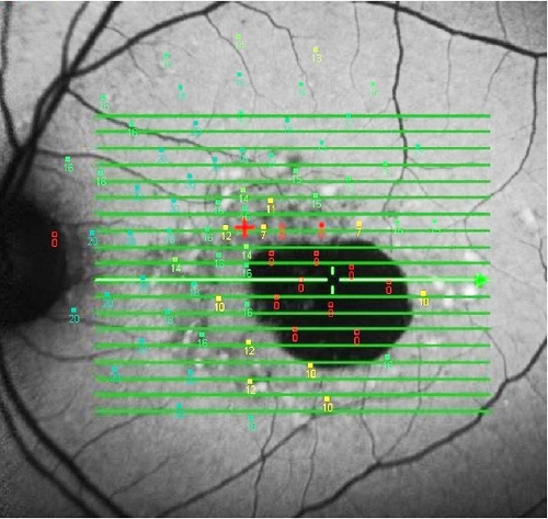
Fifteen people suffering from Stargardt’s disease were subjected to a complete eye examination and a series of diagnostic tests and procedures, such as OCT [Optical Coherence Tomography], fundus autofluorescence (FAF) and microperimetry, at the National Centre of Services and Research for the Prevention of Blindness and Visual Rehabilitation of the Visually Impaired and in the Ophthalmology Clinic of the A. Gemelli Polyclinic in Rome.
Through their careful analysis, specialists identified a significant correlation between the magnification used, the reading speed and retinal sensitivity. The latter was fundamental to assess the residual vision of the patients involved in the study, who were 42.6 years old on average and had been affected by Stargardt’s disease for at least five years.
In their scientific paper published on the Canadian Journal of Ophthalmology, the ophthalmologists and orthoptists of the National Centre and of the A. Gemelli Polyclinic concluded that:
As a result of the overlapping of OCT/FAF imaging on microperimetric exam, residual activity of outer retinal layers passing through the eccentric fixation area seems to be related with required magnification and reading rate. Identification of morpho-functional parameters is helpful for designing a customized rehabilitative program.
Useful Link: Low Vision Centers in Italy
Main Source: Can. J. Ophtalmology